human anatomy laboratory manual with cat dissections
Human anatomy laboratory manuals, often incorporating cat dissections, are crucial for students. They build understanding of biological structures through practical exploration and detailed guides.
Binkley’s guide exemplifies a resource emphasizing accuracy, safety, and educational value within feline anatomy studies.
Purpose of the Laboratory Manual
This laboratory manual serves as a comprehensive guide for students embarking on the study of human anatomy, skillfully integrated with cat dissections. Its primary purpose is to foster a deep and practical understanding of anatomical structures, moving beyond theoretical knowledge to hands-on experience.
By utilizing the cat as a comparative model, students can readily observe and appreciate the similarities and differences between feline and human anatomy. This comparative approach reinforces learning and provides a broader perspective on mammalian anatomy. The manual directs readers through detailed dissection activities, accompanied by full-color visuals, ensuring clarity and accuracy.
Furthermore, it emphasizes safety protocols and responsible dissection techniques, preparing students for future laboratory work and research endeavors. Ultimately, this manual aims to equip students with the foundational knowledge and skills necessary for success in biological sciences and related fields.
Importance of Comparative Anatomy
Comparative anatomy is fundamentally important when utilizing a human anatomy laboratory manual alongside cat dissections. Studying feline anatomy provides a valuable framework for understanding human structures, highlighting evolutionary relationships and functional similarities. Observing the cat’s skeletal and muscular systems reinforces knowledge of corresponding human systems.
This approach allows students to appreciate how anatomical structures have been modified over time to suit different lifestyles and environments. The cat, as a mammal, shares significant anatomical features with humans, making it an excellent model for comparative study.
By recognizing these similarities and differences, students gain a deeper insight into the principles of anatomy and physiology, enhancing their overall biological understanding and preparing them for advanced studies.
Safety Precautions in the Lab
When utilizing a human anatomy laboratory manual with cat dissections, strict adherence to safety protocols is paramount. Always wear appropriate personal protective equipment (PPE), including gloves and aprons, to prevent contact with biological materials. Ensure adequate ventilation in the lab and handle dissection tools with extreme care, keeping them sharp and focused on the task.
Dispose of all biological waste properly, following the instructor’s guidelines. Cover work surfaces with absorbent materials like newspapers to contain fluids. Rotate dissection responsibilities within groups to ensure everyone participates safely.
Maintain a clean and organized workspace, and immediately report any accidents or spills to the instructor. Prioritize a respectful and cautious approach throughout the dissection process.

Skeletal System
Human anatomy laboratory manuals, with cat dissections, highlight skeletal similarities. Observing feline structures reinforces understanding of human skeletal muscles and overall anatomy.
Feline vs. Human Skeletal Similarities
Human anatomy laboratory manuals utilizing cat dissections effectively demonstrate fundamental skeletal parallels. Many skeletal muscles in cats closely mirror those found in humans, providing a valuable comparative learning experience. This dissection reinforces knowledge of human skeletal muscles, allowing students to observe the protective fascia surrounding and compartmentalizing these structures.
The feline skeleton, while adapted for quadrupedal locomotion, shares a common underlying blueprint with the human skeleton. Both possess similar bone structures in the limbs, vertebral column, and skull, albeit with proportional differences reflecting their respective lifestyles. Studying these similarities enhances comprehension of mammalian skeletal anatomy and evolutionary relationships. The manual guides students to identify these shared features during dissection.
Cat Skull Anatomy
Human anatomy laboratory manuals featuring cat dissections dedicate significant attention to cranial anatomy. The cat skull, while differing in proportions from the human skull, provides an accessible model for understanding bone structure and function. Dissection reveals the various cranial bones, their sutures, and associated foramina – openings for nerves and blood vessels.
Students learn to identify key features like the orbits, nasal cavity, and temporal fossa. Understanding the feline skull’s morphology aids in comprehending the evolutionary adaptations related to sensory perception and predatory behavior. The manual guides precise identification of these structures, fostering a deeper appreciation for comparative anatomy.
Cranial Bones
Human anatomy laboratory manuals utilizing cat dissections emphasize identifying cranial bones. These include the frontal, parietal, temporal, occipital, sphenoid, and ethmoid bones. Students learn to locate sutures – the immovable joints between cranial plates – such as the sagittal and coronal sutures.
Dissection allows for tactile examination of these bones, revealing their texture and relative positions. The manual guides identification of key landmarks like the zygomatic arch and the mastoid process. Understanding the arrangement of cranial bones is fundamental to grasping brain protection and the pathways for cranial nerves.
Dental Formula & Teeth Identification
Human anatomy laboratory manuals, paired with cat dissections, detail dental formulas – a shorthand representing the number of each tooth type. Cats exhibit a dental formula of 3/3, 1/1, 3/2, 1/3, indicating incisors, canines, premolars, and molars in each jaw quadrant.
Students identify these teeth during dissection, noting their shapes and functions. Incisors are for grooming and nipping, canines for tearing, premolars and molars for grinding. Manuals highlight differences between feline and human dentition, emphasizing adaptations for carnivorous diets. Accurate teeth identification aids understanding of digestive processes and evolutionary adaptations.
Vertebral Column – Regional Differences
Human anatomy laboratory manuals utilizing cat dissections emphasize regional variations within the vertebral column. Cats possess five vertebral regions: cervical, thoracic, lumbar, sacral, and caudal. Manuals guide students to identify these regions based on vertebral morphology – shape and processes.
The cervical vertebrae are smallest, supporting the head, while thoracic vertebrae articulate with ribs. Lumbar vertebrae are larger, bearing weight. The sacral vertebrae fuse to form the sacrum, connecting to the pelvis. Finally, the caudal vertebrae form the tail. Comparing feline and human vertebral columns reveals adaptations for different locomotion styles.
Appendicular Skeleton – Limb Structure
Human anatomy laboratory manuals featuring cat dissections detail the appendicular skeleton – limbs and girdles. The feline forelimb parallels the human arm, comprising the scapula, humerus, radius, ulna, carpals, metacarpals, and phalanges. Similarly, the hindlimb corresponds to the human leg, with the pelvis, femur, tibia, fibula, tarsals, metatarsals, and phalanges.
Manuals guide students to identify these bones and observe their articulations. Cats exhibit digitigrade locomotion, walking on their toes, influencing limb bone proportions. Comparing feline and human limb structures highlights evolutionary adaptations for different modes of movement and weight distribution.

Muscular System
Human anatomy laboratory manuals with cat dissections explore muscle tissues. Observing cat muscles reinforces human skeletal muscle knowledge, examining fascia surrounding and protecting these structures.
Overview of Muscle Tissue Types
Muscle tissue, fundamental to movement, comprises several distinct types. Skeletal muscle, responsible for voluntary movements, exhibits a striated appearance under microscopy and is crucial for locomotion. Smooth muscle, found in the walls of internal organs, controls involuntary functions like digestion.
Cardiac muscle, exclusive to the heart, possesses unique structural and functional characteristics enabling rhythmic contractions. A human anatomy laboratory manual utilizing cat dissections allows students to directly observe these tissue types. Comparing feline and human muscle structures enhances understanding of evolutionary relationships and functional adaptations. Dissection reveals muscle fascicles and fiber arrangements, illustrating how these elements contribute to muscle force and range of motion.
Students can identify and differentiate these tissues, solidifying their grasp of muscular system physiology.
Superficial Muscles of the Cat – Identification
Cat dissections, guided by a human anatomy laboratory manual, facilitate identification of superficial muscles. Key muscles include the platysma, a thin sheet covering the neck, and the omohyoid, supporting the hyoid bone. The sternocleidomastoid, prominent on the sides of the neck, enables head movement.
Students observe the pectoralis, responsible for limb protraction, and the latissimus dorsi, aiding in limb extension. Identifying the external abdominal oblique and rectus abdominis reveals core musculature. Comparing these to homologous human muscles reinforces anatomical knowledge. Observing fascia surrounding these muscles demonstrates compartmentalization and protection.
Careful dissection and accurate labeling are essential for mastering feline superficial anatomy.
Deep Muscles of the Cat – Identification
Utilizing a human anatomy laboratory manual during cat dissection allows for identification of deep muscles. These lie beneath superficial layers, requiring careful dissection. Key muscles include the splenius, extending the head and neck, and the longissimus dorsi, contributing to vertebral column stability.
Students can identify the iliocostalis, part of the erector spinae group, and the transversus abdominis, crucial for core support. Observing the psoas major and iliacus reveals hip flexor anatomy. Comparing these to human counterparts highlights evolutionary similarities.
Precise identification and understanding of muscle origins and insertions are vital for a comprehensive anatomical study.
Muscle Fascicles and Fiber Arrangement
A human anatomy laboratory manual, paired with cat dissection, reveals muscle organization. Muscles aren’t uniform; they’re composed of fascicles – bundles of muscle fibers. Fiber arrangement dictates a muscle’s strength and range of motion.
Parallel arrangements, like in the sartorius, prioritize range. Pennate arrangements, such as in the rectus femoris, maximize strength. Observing the angle of pennation is key. Fusiform muscles, wider at the belly, demonstrate a balance.
Understanding fascicle structure clarifies how muscles generate force and movement. Dissection reveals the protective fascia surrounding these bundles, compartmentalizing and supporting muscle function.

Nervous System
Human anatomy laboratory manuals, utilizing cat dissections, illuminate neurological structures. Comparative study reveals similarities in brain anatomy, spinal cords, and peripheral nerve distribution.
Brain Anatomy – Comparative Overview
Comparative brain anatomy, facilitated by human anatomy laboratory manuals and cat dissections, reveals striking parallels. Both species exhibit a similar tripartite structure: cerebrum, cerebellum, and brainstem. The cerebrum, responsible for higher cognitive functions, demonstrates gyri and sulci, though proportionally larger in humans.
The cerebellum, crucial for motor coordination, appears relatively consistent in both, though subtle differences exist in lobation. The brainstem, governing vital functions, showcases homologous structures like the midbrain, pons, and medulla oblongata. Dissection allows visualization of these regions, enhancing understanding of functional localization.
Students can trace nerve pathways and appreciate the evolutionary conservation of brain organization, solidifying anatomical knowledge through hands-on exploration.
Cerebrum & Lobes
The cerebrum, the largest brain region, exhibits distinct lobes – frontal, parietal, temporal, and occipital – observable in both humans and cats through dissection guided by human anatomy laboratory manuals. While the overall structure is conserved, the degree of cortical folding (gyrification) differs significantly.
Humans possess a highly convoluted cerebrum, maximizing surface area for complex cognitive processing. Cats exhibit less gyri, reflecting their comparatively simpler cognitive demands. Identifying these lobes during dissection reinforces understanding of their respective functions: motor control (frontal), sensory integration (parietal), auditory processing (temporal), and visual processing (occipital).
Comparative analysis highlights evolutionary adaptations in brain structure relating to behavioral complexity.
Cerebellum & Function
The cerebellum, a crucial structure for motor control, coordination, and balance, is readily identifiable during cat dissection using a human anatomy laboratory manual. Though smaller relative to the cerebrum in both species, its distinct lobulation and consistent location posterior to the brainstem are key features.
Comparative observation reveals similarities in cerebellar structure, supporting its conserved role in fine-tuning movements. However, humans exhibit a more developed cerebellum, correlating with their capacity for complex motor skills. Dissection allows tracing cerebellar peduncles – fiber bundles connecting it to the brainstem – vital for information transfer.
Understanding cerebellar function enhances appreciation for coordinated movement.
Spinal Cord & Nerve Roots
Utilizing a human anatomy laboratory manual alongside cat dissection provides a valuable opportunity to study the spinal cord and its associated nerve roots. The spinal cord, a cylindrical structure extending from the brainstem, transmits signals between the brain and the periphery.
Dissection reveals the dorsal and ventral roots, formed by sensory and motor neurons respectively, converging to create spinal nerves. Observing these structures in the cat mirrors human anatomy, highlighting the fundamental organization of the central nervous system. Identifying the grey and white matter distribution within the cord is also crucial.
This comparative approach reinforces understanding of neurological pathways.
Peripheral Nervous System – Major Nerves
A human anatomy laboratory manual, complemented by cat dissection, allows for detailed examination of the peripheral nervous system and its major nerves. These nerves extend from the central nervous system, branching throughout the body to innervate muscles and sensory receptors.
Dissection reveals key nerves like the brachial plexus (forelimb innervation) and lumbar plexus (hindlimb innervation). Tracing these nerves demonstrates their pathways and the regions they serve. Comparing feline and human nerve distribution highlights conserved anatomical patterns.
Identifying nerve origins and branches enhances understanding of motor control and sensory perception.

Digestive System
Human anatomy laboratory manuals, utilizing cat dissections, illuminate the digestive tract’s structure. Exploration reveals organs from the oral cavity to the rectum, aiding comparative analysis.
Oral Cavity & Accessory Organs
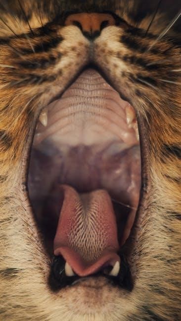
Human anatomy laboratory manuals, enhanced by cat dissections, provide detailed insights into the oral cavity and its associated structures. Dissection allows for direct observation of the feline mouth, revealing similarities and differences compared to human anatomy.
Students can identify key components like the teeth, tongue, and hard/soft palates. Examining accessory organs – salivary glands – demonstrates their role in initial food processing. Comparative analysis highlights variations in dental formulas and gland morphology between species.
This practical experience reinforces understanding of digestive function and evolutionary adaptations. Careful dissection and identification of structures are essential for grasping the complexities of the digestive system in both humans and cats.
Esophagus & Stomach Anatomy
Human anatomy laboratory manuals utilizing cat dissections offer a comparative view of the esophagus and stomach. Dissection reveals the muscular tube of the esophagus, responsible for peristaltic transport of food. Observing the feline esophagus aids in understanding its human counterpart’s structure and function.
The stomach, a J-shaped organ, is examined for its layers – mucosa, submucosa, muscularis externa, and serosa. Students identify the cardia, fundus, body, and pylorus. Comparing feline and human stomach anatomy highlights differences in size and shape, reflecting dietary habits;
This hands-on experience solidifies knowledge of digestive processes and anatomical variations, crucial for a comprehensive understanding of the mammalian digestive system.
Small Intestine – Duodenum, Jejunum, Ileum
Human anatomy laboratory manuals, enhanced by cat dissections, provide detailed study of the small intestine’s three segments. The duodenum, receiving chyme from the stomach, is identified by its C-shape and bile duct entry. The jejunum, with its prominent villi, demonstrates nutrient absorption’s primary site.
The ileum, the final segment, connects to the large intestine and features Peyer’s patches for immune surveillance. Comparing feline and human small intestine lengths and structural features reveals adaptations to different diets.
Dissection emphasizes the intestinal wall’s layers and the importance of villi and microvilli in maximizing surface area for efficient nutrient uptake, solidifying digestive physiology understanding.
Large Intestine – Cecum, Colon, Rectum
Human anatomy laboratory manuals utilizing cat dissections illuminate the large intestine’s structure. The cecum, a blind pouch, is comparatively smaller in cats than humans, reflecting dietary differences. The colon – ascending, transverse, and descending – demonstrates water absorption and waste compaction.
Dissection reveals haustra, sacculations aiding in colonic movement. The rectum stores feces before elimination via the anal canal. Comparing feline and human colonic lengths and the presence of a distinct cecum highlights evolutionary adaptations.
Observing the intestinal wall’s muscular layers and the absence of villi reinforces understanding of its primary function: water recovery and waste preparation for excretion.

Respiratory System
Human anatomy laboratory manuals, with cat dissections, detail the respiratory pathway. Examining the nasal cavity, larynx, trachea, lungs, and bronchial tree provides comparative insights.
Nasal Cavity & Pharynx
Human anatomy laboratory manuals utilizing cat dissections allow for a comparative study of the nasal cavity and pharynx. In both species, the nasal cavity functions to warm, humidify, and filter incoming air before it reaches the lungs. Dissection reveals the turbinates, bony structures increasing surface area within the nasal passages.
The pharynx, a shared pathway for both air and food, demonstrates structural similarities. Students can observe the nasopharynx, oropharynx, and laryngopharynx in the cat, correlating these regions to human anatomy. Careful dissection highlights the epiglottis, crucial for preventing aspiration during swallowing. Understanding these structures in the cat provides a valuable foundation for comprehending their function in humans.
Comparative analysis emphasizes evolutionary relationships and functional adaptations within the respiratory system.
Larynx & Trachea
Human anatomy laboratory manuals employing cat dissections facilitate the study of the larynx and trachea, vital components of the respiratory system. The larynx, or voice box, contains vocal cords responsible for sound production; dissection reveals cartilaginous structures like the thyroid and cricoid cartilages.
The trachea, a flexible tube reinforced by C-shaped cartilage rings, maintains an open airway. Students observe the tracheal rings during dissection, understanding their role in preventing collapse. Comparing feline and human tracheal anatomy highlights similarities in structure and function.
These practical observations reinforce theoretical knowledge, solidifying understanding of respiratory mechanics and anatomical relationships. Careful dissection aids in identifying key landmarks and appreciating the system’s overall design.
Lungs – Lobes & Bronchial Tree
Human anatomy laboratory manuals utilizing cat dissections provide invaluable insight into lung structure. The feline lung exhibits distinct lobes – typically three on the right and two on the left – observable during dissection. Students identify these lobes and trace the branching pattern of the bronchial tree.
The bronchial tree, starting with the primary bronchi, progressively divides into smaller bronchioles, culminating in alveoli, the sites of gas exchange. Dissection allows visualization of this hierarchical structure, demonstrating increasing surface area.
Comparing feline and human lung lobation reveals anatomical differences. This practical experience enhances comprehension of respiratory physiology and the importance of lung architecture.
Cardiovascular System
Human anatomy laboratory manuals, with cat dissections, reveal heart chambers and vessels. Students trace major arteries and veins, comparing feline and human circulatory systems.
Heart Anatomy – Chambers & Vessels
Detailed dissection within a human anatomy laboratory manual, utilizing cat dissections, allows for precise identification of the heart’s four chambers: the right and left atria, and the right and left ventricles. Students observe the differing thicknesses of ventricular walls, correlating to pulmonary versus systemic circulation.
Key vessels, including the superior and inferior vena cava, pulmonary artery, and aorta, are traced. The manual guides students to locate the coronary arteries and veins, understanding their vital role in cardiac muscle perfusion. Careful observation reveals the atrioventricular and semilunar valves, crucial for unidirectional blood flow.
Comparative analysis highlights similarities and differences between feline and human cardiac anatomy, reinforcing understanding of mammalian cardiovascular function.
Major Arteries & Veins
A human anatomy laboratory manual, enhanced by cat dissections, facilitates tracing the circulatory pathways. Students identify major arteries – aorta, carotid, subclavian – noting their branching patterns and destinations. The manual guides observation of the arterial system supplying limbs and vital organs.
Corresponding veins, including the vena cava (superior and inferior), jugular, and subclavian, are located and their roles in returning deoxygenated blood to the heart are understood. The manual emphasizes identifying the femoral artery and vein in the cat’s hindlimb.
Comparative anatomy reveals conserved patterns, while highlighting species-specific variations in vessel size and distribution, solidifying comprehension of mammalian circulatory systems.

Urogenital System
Human anatomy laboratory manuals, utilizing cat dissections, detail kidney structure, bladder function, and reproductive organs. Comparative analysis reveals similarities and distinctions.
Kidneys & Urinary Bladder
Cat dissection manuals, alongside human anatomy studies, emphasize the kidney’s role in filtration and waste removal. Observe the feline kidney’s cortical and medullary regions, comparing them to human structures. Note the renal artery and vein, crucial for blood supply.
The urinary bladder’s function in urine storage is also examined. Dissection reveals its muscular walls and connection to the urethra. Compare the relative sizes and positions of these organs in cats versus humans.
Laboratory exercises often involve tracing the urinary pathways, reinforcing understanding of the urogenital system’s integrated function. Detailed illustrations within manuals aid in accurate identification of anatomical features.
Male Reproductive System
Cat dissection, within a human anatomy context, allows for comparative study of the male reproductive organs. Identify the testes, located within the scrotum, and trace the epididymis and vas deferens. Observe the seminal vesicles and prostate gland, crucial for fluid production.
Manuals guide students through locating the penis and its associated structures. Compare the feline penis’s unique features, like barbed spines, to the human anatomy. Note the position of the urethral opening.
Understanding the pathway of sperm is key. Dissection reinforces knowledge of hormone production and the overall function of the male reproductive system in both species.
Female Reproductive System
Cat dissection, alongside a human anatomy lab manual, facilitates comparative study of the female reproductive tract. Locate the ovaries, often small and difficult to find, and trace the fallopian tubes (oviducts) leading to the uterine horns.
Identify the uterus, which is bicornuate in cats – meaning it has two distinct horns. Observe the cervix, connecting the uterus to the vagina. Manuals aid in visualizing these structures.
Compare the feline estrous cycle to the human menstrual cycle. Note the absence of a distinct endometrium in non-pregnant cats. Dissection reinforces understanding of hormone regulation and reproductive function in both species.